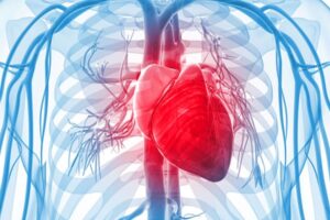
Cardiac or heart abnormalities are often associated with Noonan syndrome. These can include problems with the heart valves, holes in the walls separating the chambers of the heart, and thickening of the heart muscle.
People with Noonan syndrome can be affected by a wide range of heart-related disease, which fall into two main categories:
Congenital heart disease affects about 80% of patients
Thickened heart muscle (hypertrophic cardiomyopathy) is found in about 20% of patients.
Heart problems in Noonan syndrome
Common:
Pulmonary valve stenosis The pulmonary valve (the valve between the right ventricle and the pulmonary artery that goes to the lungs) is too small, narrow, or stiff.
Moderate risk:
Hypertrophic cardiomyopathy The heart muscle becomes abnormally thick (hypertrophied).
Rare:
Atrial septal defect A hole in the wall (septum) that divides the upper chambers (atria) of the heart.
Ventricular septal defect A hole in the wall (septum) that separates the two lower chambers (ventricles) of the heart Atrioventricular septal defect A large defect in the centre of the heart that can be a combination of:
atrial septal defect
ventricular septal defect
abnormalities of the valves, often resulting in one large valve instead of two separate valves
Mitral abnormalities A problem with the valve located between the left heart chambers (left atrium and left ventricle)
Aortic coarctation Part of the aorta – the large artery that carries oxygen-rich blood to the rest of the body – is narrower than usual.
Patent ductus arteriosus A persistent opening between the two major blood vessels leading from the heart. The opening, a normal part of a baby’s circulatory system in the womb, usually closes shortly after birth.
Tetralogy of Fallot Complex abnormality with four defects of the heart:
Ventricular septal defect;
Pulmonary stenosis;
Enlarged and abnormal aortic valve;
Ventricular hypertrophy – the muscular wall of the lower right chamber of the heart (right ventricle) is thicker than normal.
Pulmonary valve stenosis
Affecting about 40% of people with Noonan syndrome, pulmonary valve stenosis is the most common heart problem. It occurs when the pulmonary valve (the valve between the right ventricle and the pulmonary artery that goes to the lungs) is too small, narrow, or stiff.
The severity varies:
Mild cases (about 60%): these cases are similar to patients with pulmonary valve stenosis who do not have Noonan syndrome – the stenosis tends not to progress (and often improves without treatment) and may not need surgical treatment. However, in some cases there may be an additional heart problem such as an atrial septal defect.
Moderate (about 10%) or severe (about 30%) cases: in these cases, the rates of surgical treatment are higher – about 50% and 100%, respectively.
Atrioventricular septal defects
An atrioventricular septal defect (AVSD) is a large defect in the centre of the heart that can be a combination of:
atrial septal defect
ventricular septal defect
abnormalities of the valves, often resulting in one large valve instead of two separate valves
A partial atrioventricular septal defect is the most common form of this problem, but complete defects, while rare, do occur.
Hypertrophic cardiomyopathy
About 10 – 20% of people with Noonan syndrome suffer from hypertrophic cardiomyopathy, where the heart muscle becomes abnormally thick (hypertrophied). This can become evident early in life, with more than half diagnosed by six months of age (much earlier than other childhood forms of hypertrophic cardiomyopathy, which on average become evident at the age of 8 years). Obstructions of the outflow from the left ventricle are common, and children with Noonan syndrome and hypertrophic cardiomyopathy are more likely to have congestive heart failure than other children with hypertrophic cardiomyopathy.
Genetics
A mutation in the PTPN11 gene is the most common cause of Noonan syndrome but there are now up to 15 genes which may be involved. The gene affected can also influence the likelihood and type of congenital heart disease.
PTPN11 mutation
More likely to have pulmonary valve stenosis or an atrial septal defect
Less likely to have hypertrophic cardiomyopathy
RAF1 mutation
Less likely to have pulmonary valve stenosis
More likely to have hypertrophic cardiomyopathy
RIT1 mutation
More likely to have hypertrophic cardiomyopathy and valve abnormalities
SOS1 mutation
More likely to have pulmonary valve stenosis
Treatment
About a third of patients with pulmonary stenosis needed surgery or a repeat procedure.
A balloon can be used to open a stiff valve, through a procedure termed ‘percutaneous balloon valvuloplasty’. If this fails or is deemed not feasible, surgical valvotomy – an open-heart procedure to open up a valve – can be carried out.
Other structural heart defects may require invasive treatment. Some may be amenable to catheter intervention (e.g. percutaneous balloon valvuloplasty, stent insertion or device closure) but others may require cardiac surgery. Treatment of other structural heart defects – these may require cardiac surgery
Treatment of hypertrophic cardiomyopathy is primarily directed at treating symptoms. This is usually with medication (e.g. beta blockers), although rarely cardiac surgery to relieve left ventricular outflow tract obstruction or to repair or replace the mitral valve may be required. Very rarely, individuals with Noonan syndrome and hypertrophic cardiomyopathy may be at an increased risk of potentially life-threatening abnormal heart rhythms and treatment with an implantable cardioverter-defibrillator (ICD) may be recommended. More recently, a new class of drugs (MEK inhibitors such as trametinib) has been reported to improve the features and symptoms of hypertrophic cardiomyopathy in some children with Noonan syndrome and related conditions presenting with very severe forms in the first few months of life. Further studies to investigate the use of these drugs are ongoing.
References
Linglart L, Gelb BD. Congenital heart defects in Noonan syndrome: Diagnosis, management, and treatment. Am J Med Genet C Semin Med Genet. 2020;184(1):73-80.
Burch M, Sharland M, Shinebourne E, Smith G, Patton M, McKenna W. Cardiologic abnormalities in Noonan syndrome: phenotypic diagnosis and echocardiographic assessment of 118 patients. J Am Coll Cardiol. 1993 Oct;22(4):1189-92.
Roberts AE, Allanson JE, Tartaglia M, Gelb BD. Noonan syndrome. Lancet. 2013;381(9863):333-342.
Gelb BD, Roberts AE, Tartaglia M. Cardiomyopathies in Noonan syndrome and the other RASopathies. Prog Pediatr Cardiol. 2015;39(1):13-19.
I am a 56-year-old adult male with Noonan Syndrome. What, if any, effect might the condition have on me as I get older?
In general terms, the main issue is the narrowing of the pulmonary valve in the heart in childhood which may recur again after treatment later in life. There are a number of rarer complications that may occur, but as you have mentioned no symptoms they are unlikely. The rare complications include swelling of the legs, problems with the gut, weight loss, and disturbances in the heart pumping or heart rhythm.
Can my daughter who has NS (PTPN11) and HC have her ears pierced?
I would seek the advice of her cardiologist. In our practice, HCM is not a contraindication to ear piercings but there may be other heart lesions (e.g. leaky valves) that may mean the cardiologist would want to be a bit more cautious.
Our daughter suffers from spontaneous chylothorax as well as pulmonary stenosis and ASD. She’s had open heart surgery, which was very successful. I was wondering if in the medical world there is any more research being done on whether there is any connection to the chylothorax issues within her lungs to cardiac issues, or if they are totally separate situations?
There’s certainly association with chylothorax and cardiac surgery with individuals who have had open heart surgery who then develop chylothorax. We also know there is an increased prevalence of lymphatic problems in Noonan Syndrome. There is a lot of research going on in terms of the work Sahar is doing and the fact that we can now image lymphatics in a better way. We’ve talked about Noonan’s having some implications for growth and potentially over-growth, over-thickening of vessels. There’s a possibility that certain situations lead to over-growth of lymphatic vessels which then don’t do their job very well. There’s an excellent link now with Philadelphia with Professor Max Itkin who is an expert in interventional radiology to actually get a picture of the whole lymphatics of the body, not just the lower part of the body and then ways to intervene in certain circumstances.
What research is going on for adults aged over 30 with Noonan Syndrome, and where can they get help and advice?
A study with a cohort of families started in 1985 and most of them are now young adults in their twenties. Hopefully the group can continue to be followed up so that we’ll really know what’s happening later in life. There was also a collaborative effort across Europe to try to understand the natural history of the cardiac complications of Noonan Syndrome. Also, clinically, there are a number of specialised centres for example cardiomyopathy expert centres in the UK, which have a lot of experience of looking after adults with Noonan Syndrome-related congenital heart disease. It is important to remember however that Noonan Syndrome was not progressive. There could be complications later in life but many people remain in good health with no further problems.
Would the growth hormone be risky for someone who has RAF1, which is high risk for HOCM, but doesn’t have it yet?
We currently do not have enough information to know whether growth hormone is riskier in certain genes, but the risk is more likely to be related to whether there is already significant HCM rather than the gene itself. We are still collecting information on this, though.
What are the chances of my child with pulmonary stenosis, who has had open heart surgery in the past, having more surgery as she gets older?
It’s very variable. It depends on the initial condition and the initial procedure performed. It’s not unusual for individuals who’ve had particularly surgery to the pulmonary valve for that valve in time to become leaky and often that does require further procedure. It’s not universal by any means and it depends on the shape and the look of the valve – the morphology of the valve and the procedure that was performed. It is well established that anyone who’s had an intervention to their pulmonary valve should have lifelong follow up. There are adult congenital heart services around the country which would provide that sort of follow up.
A lot of NS children have had open heart surgery for pulmonary stenosis. Will future developments in technique mean a less invasive treatment?
Less invasive techniques were available for more straightforward cases to open up a pulmonary valve but this “balloon” procedure didn’t work as well when the valve was very thick or abnormally formed. In these cases, surgeons often choose to go for open heart surgery as the result is likely to be better first-time round. But in many individual cases of children with NS it was possible to do the less invasive balloon dilation technique and hopefully that these techniques will improve in the future.
My son bruises very easily when he falls over and the bruises last a very long time. He’s unfortunately got both pulmonary and aortic valve problems so he’ll need an artificial valve when his heart deteriorates to a certain level and then he’ll have to go on blood thinners. What considerations would there need to be regarding his bruising issues?
There has been a fairly large study of clotting factors in children with Noonan’s which found that there were occasional issues with factor 8 clotting agent but usually these were not causing serious bleeding although they did cause bruising more easily. In rare cases of post-operative bleeding, it was relatively easy to correct that with fresh blood which did contain the clotting agent. If it did come to an operation for the child in question, the specific needs would be looked at carefully and there were options as regards blood thinners as new ones were being developed, although very few were licenced for use with children at the moment. There are artificial valves which don’t require anti-coagulation but as in most cases, it very much depended on the individual case and there would be very detailed investigation before a decision was made. There had however been several cases where Warfarin had been used with children with Noonan’s.
Our daughter has moderate pulmonary stenosis. She is nearly 3. Our son is 4 (both Noonan’s LZTR1). No detected cardiac issues. Is there any indication of the age any other cardiac issues—if they occur—might become apparent?
We are still learning about how the heart is affected in LZTR1, but it is very unlikely that any valve changes will develop if they are not present now. What can develop over time, and needs regular follow up, is hypertrophic cardiomyopathy. In most cases, we think this is often present very early on, but it can sometimes develop later in life, so we would recommend ongoing cardiac screening.
My son, 29, teacher with Tetralogy of Fallot, has a leaky pulmonary valve—is he likely to need further surgery as he gets older?
In some cases, after repair of tetralogy of Fallot, the pulmonary valve can become leaky, and this sometimes does need further surgery. It depends on the technique used for the original surgery, the degree of leakiness and the effect the leakiness has on the right side of the heart.
My son has RAF1, and he now has a moderate leaky mitral valve which was trivial and changed within 6 months to moderate along with a new leaky aortic valve. Can they self-correct or do they deteriorate, needing surgery? My son is 10 years old.
Everybody is different, and in some cases, the amount of leakiness can progress to the extent that it causes symptoms and needs surgery; in other cases, the leakiness can stay stable for many years and not cause any problems. The key is to make sure that he remains under close follow up.
My son has RAF1, and we now know we all have this same mutation. His cardiologist mainly checks for HOCM, however nobody in this family has HOCM. My son has a leaky aortic valve and a leaking mitral valve. Could the RAF1 diagnosis be incorrect, as RAF1 is associated with HOCM and not valve leakages?
Although RAF1 variants are more commonly associated with HCM, not everyone with RAF1 always has HCM, and there is a lot of overlap with other conditions, such as leaky heart valves. It is likely the RAF1 diagnosis is correct, but that is hasn’t caused HCM in the family. He should remain under follow up, though, as sometimes the HCM develops later.
My fifteen-year-old granddaughter has hypertrophic cardiomyopathy and has very heavy periods. Can she be given progesterone or the contraceptive pill to help with the period problems taking into account the hypertrophic cardiomyopathy?
This would need to be checked with her own cardiologist. It’s true that there are certain heart conditions where the oral contraceptive pill is contra-indicated and that’s because in some situations there’s an increased risk of stroke. There are some other treatments for very heavy periods that may be considered but again you may have to be guided by specialists such as a gynaecologist.
Is dysplastic aortic valve seen very often in Noonan Syndrome and what are the potential symptoms and interventions for this?
Although less common than pulmonary valve dysplasia, it is not unusual to see dysplasia of the other valves, including the aortic valve, in Noonan Syndrome. A dysplastic valve on its own shouldn’t cause any symptoms, but if it becomes narrowed or leaky, it may do, and that may require intervention (keyhole or surgical) if there is significant narrowing or leakiness.
Can there be a risk of developing heart issues later in life if there are none at the moment?
Not usually. You may want to check out our webinar on heart issues in NS.
Can HCM cause sudden death? Are there any warning signs to look out for?
In a small number of patients, HCM can cause sudden death, although this is much rarer in Noonan syndrome/RASopathies than in other forms of HCM. We have found that certain markers on echo and the history can help identify which patients with HCM have a higher risk and would benefit from implantation of an ICD – the cardiologist will be looking at these markers and will be best placed to decide whether there are any markers of increased risk
Are respiratory problems quite common with Noonan Syndrome? My daughter has pulmonary stenosis, HCM and an ASD but everything that keeps putting her in hospital is respiratory. Any infection turns into a major infection on her chest and hospitalises her. She’s been admitted thirty-two times in two years.
It’s sometimes very difficult to distinguish lung related causes and heart related causes in this situation and we also know that respiratory infections that might be very well tolerated in individuals without heart disease often are much less well tolerated in children who have cardiac involvement, particularly when there’s a combination of things. So if you have hypertrophic cardiomyopathy and pulmonary valve stenosis and an ASD, these are three different things that effect the heart in slightly different ways and so we’ve certainly seen that children who present with chest infections that you wouldn’t normally expect to cause significant symptoms, take a lot longer to get better, and often children feel much worse with them during that time. I’m sure that they’re right that the cause of the deterioration is respiratory – a lung related issue but I’m sure that the heart is contributing in that it’s making things worse. Individuals with a cardiac condition are eligible for an immunisation for the RSV virus which is worth exploring. Another thing to think about here is family history of respiratory conditions. For a child to be admitted thirty-two times is quite marked, there’s something going on there that does need to be evaluated. There could be some immunological investigations that could be helpful. It may also be a consequence of small micro-aspirations that she has now developed a condition whereby she is oxygen dependent. Again, its things we can address and she could grow out of but we need to know the causes so there will be all sorts of tests like speech, language and swallow assessments.
We use tools which employ cookies to collect anonymous information about how visitors use our site.
If you are not happy for us to monitor your use of our website, and you do not wish to disable cookies on your browser, then do not use our website. OK